


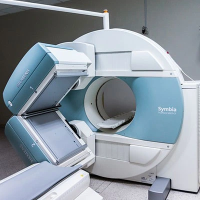
Description
Test Preparation
To ensure accurate results, follow these preparation guideline:
• Fasting: Usually not required, but if contrast dye is used, fasting for 4-6 hours may be recommended.
• Clothing & Accessories: Wear loose, comfortable clothing without metal (zippers, buttons, jewelry). A hospital gown may be provided.
• Metal Objects: Remove glasses, dentures, piercings, hearing aids, or hairpins as MRI uses a strong magnet.
• Medical Implants: Inform the radiologist if you have pacemakers, aneurysm clips, cochlear implants, or metallic implants, as MRI may not be safe.
• Claustrophobia: If you feel claustrophobic (fear of enclosed spaces), let your doctor know. A mild sedative may be prescribed.
• Pregnancy & Breastfeeding: Inform your doctor if you are pregnant or breastfeeding, especially if contrast dye is needed.
• Contrast Dye (if applicable): If contrast is used, inform the technician about any history of allergies, kidney disease, or asthma.

