


Description
Test Preparation
To ensure accurate results, follow these preparation guideline:
• Fasting: Not typically required, but follow your doctor’s instructions.
• Medications: Continue regular medications unless instructed otherwise. If you take blood thinners, consult your doctor.
• Clothing: Wear loose, metal-free clothing. You may be asked to wear a hospital gown.
• Allergy Check: Inform your doctor if you have a history of allergies to contrast dye or kidney issues.
• Pregnancy/Breastfeeding: If pregnant or breastfeeding, discuss with your doctor whether the test is necessary.
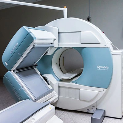
• After contrast injection, you will move to the MRI scanner.
• The scan lasts 30–60 minutes, and you must remain still.
• You may hear loud noises from the MRI machine; earplugs or headphones are provided.
• You may be asked to move your wrist slightly to assess joint function.

