


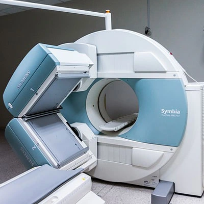
Description
Test Preparation
To ensure accurate results, follow these preparation guideline:
Clothing & Accessories:
Wear comfortable, metal-free clothing (avoid zippers, buttons, or snaps).
Remove jewelry, piercings, watches, and metallic accessories before the scan.
Medical History& Implants:
Inform the technician if you have metal implants, pacemakers, artificial joints, or aneurysm clips, as they may interfere with the MRI.
If you have kidney disease, notify them if a contrast dye is required.
Fasting (If Contrast is Required):
If a contrast dye (gadolinium) is needed, you may be asked to avoid eating or drinking for 4–6 hours before the scan.
Pregnancy & Claustrophobia:
Inform the radiologist if you are pregnant or breastfeeding.
If you have claustrophobia, discuss sedation or open MRI options with your doctor.
Medications:
Continue taking your regular medications, unless instructed otherwise by your doctor.
Arrival & Procedure:
Arrive 30 minutes before your appointment to complete paperwork.
The scan typically takes 30–60 minutes, during which you must stay still for clear imaging.

