


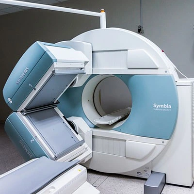
Description
Test Preparation
To ensure accurate results, follow these preparation guideline:
- General Preparation:
• Remove metal objects (jewelry, piercings, watches, eyeglasses, etc.) to prevent interference with the MRI.
• Wear comfortable, loose-fitting clothing without metal zippers, buttons, or hooks. A hospital gown may be provided.
• Inform your doctor if you have any metal implants, pacemakers, surgical clips, or neurostimulators, as these may be affected by the magnetic field. - Fasting & Medication:
• Fasting is usually not required, but if a contrast-enhanced MRI is ordered, you may need to fast for 4–6 hours before the test.
• Inform your doctor if you have any kidney disease, diabetes, or allergies, as these may affect the use of contrast dye. - Contrast Injection (if required):
• A contrast agent (gadolinium dye) may be injected through an IV for better visualization of nerves and blood vessels.
• If you have a history of allergic reactions to contrast dye, notify your doctor.
Sedation (if needed):
• If you have claustrophobia or difficulty lying still for long periods, mild sedation may be an option. - During the MRI:
• The scan takes 30–60 minutes. You must lie still for clear imaging.
• Your head, neck, and upper chest will be inside the MRI machine.
• The machine produces loud knocking sounds; earplugs or headphones may be provided for comfort.

