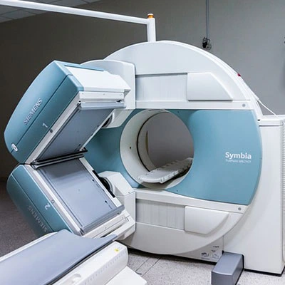
MRI Arthrogram With Shoulder

18,000.00

Categories: ,

An MRI Arthrogram of the Shoulder is a specialized imaging test used to evaluate the shoulder joint in greater detail. Unlike a standard MRI, an arthrogram involves injecting a contrast dye (gadolinium) directly into the shoulder joint before the MRI scan. This contrast helps highlight soft tissues, cartilage, ligaments, and joint structures, making it easier to detect injuries or abnormalities.

Why is it Done?
An MRI arthrogram of the shoulder is commonly performed to diagnose:
• Rotator cuff tears (including small or partial tears)
• Labral tears (e.g., SLAP tears, Bankart lesions)
• Cartilage damage (chondral injuries)
• Shoulder instability or dislocations
• Biceps tendon injuries
• Post-surgical complications

Enquiry Here

    Description

    Test Preparation

    To ensure accurate results, follow these preparation guideline:

    • Fasting: Not typically required, but follow your doctor’s instructions.
    • Medications: Continue your regular medications unless instructed otherwise. If you take blood thinners, consult your doctor as you may need to pause them before the procedure.
    • Clothing: Wear loose, comfortable clothing without metal (zippers, jewelry, or underwire bras). A hospital gown may be provided.
    • Allergy Check: Inform your doctor if you have allergies to contrast dye or a history of kidney disease.
    • Pregnancy/Breastfeeding: If you are pregnant or breastfeeding, discuss with your doctor whether the test is necessary