
Description
Test Preparation
To ensure accurate results, follow these preparation guideline:
- Fasting
• Usually, no fasting is required unless contrast dye is used.
• If contrast is needed, some facilities may require 4-6 hours of fasting. - Hydration
• Drink plenty of water before the test unless instructed otherwise. - Medications
• Take your regular medications unless advised otherwise.
• Inform your doctor if you are taking blood thinners or have kidney problems (if contrast dye is used). - Allergies & Medical Conditions
• Inform the technician if you have a history of contrast dye allergies.
• If you have kidney disease, contrast dye may require special precautions. - Clothing & Accessories
• Wear loose, comfortable clothing without metal (zippers, buttons, jewelry, or underwires).
• You may be asked to change into a hospital gown. - Implant or Device Precautions
• Inform the technician if you have metal implants, pacemakers, cochlear implants, aneurysm clips, or any metallic objects in your body, as they may interfere with the MRI. - Claustrophobia & Anxiety
• If you have claustrophobia, discuss with your doctor about a mild sedative before the test.
• Some facilities offer open MRI options for comfort. - During the Procedure
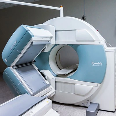
• You will lie on a table that slides into the MRI scanner.
• If contrast dye is needed, it will be injected into a vein (usually in the arm) to enhance image clarity.
• The machine makes loud thumping or knocking sounds; earplugs or headphones may be provided.
• The scan typically takes 30-60 minutes. - After the Test
• No downtime; you can resume normal activities unless sedated.
• Drink plenty of water to help eliminate the contrast dye from your system.
• Results are usually available within a few days.